| Walaikum Salam Sister Laila, welcome to the site!
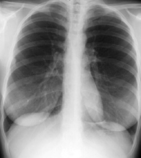
You have asked a good question, If you mean her chest X-ray which was done for pneumonia, NO, A fibroadenoma is usually diagnosed through clinical examination, ultrasound, mammography (X-ray for the breast) and often a biopsy sample of the lump.
On chest X-ray, you can see the shape, contour, regularity of the breast (breast cancer), if there is missing breast (mastectomy) or if there is silicon planted.
On examination, fibroadenomas are oval, freely mobile, rubbery masses, not tender, not fixed to the adjacent skin, muscle, or lymph nodes, so they are mobile within the breast on palpation. It is commonly found immediately adjacent to the areola, though rarely directly behind the nipple.
Imaging:
On mammograms, fibroadenomas typically appear as circumscribed oval or round masses, which occasionally have coarse calcifications.
BUT Mammography cannot be used to distinguish whether a mass is a fibroadenoma, a cyst, or a carcinoma with certainty because of some overlap in the findings. All of the entities may appear as smooth masses.
On ultrasonograms, fibroadenomas appear as circumscribed, homogeneous, oval, hypoechoic masses that may have gentle lobulations; a smooth, thin, echogenic capsule; variable acoustic enhancement; and homogeneity.
BUT On ultrasonograms, fibroadenomas often demonstrate a typical appearance and may be distinguished clearly from cysts and carcinomas; however, fibrocystic disease with complicated hypoechoic cysts and, rarely, smooth carcinomas may mimic fibroadenoma. Atypical fibroadenomas, which are inhomogeneous or irregular in shape, may simulate carcinomas.
On MRIs, fibroadenomas typically appear as smooth masses with high signal intensity on T2-weighted images and enhancement with the administration of gadolinium-based contrast agent.
BUT On MRIs, enhancement characteristics may help distinguish fibroadenomas from carcinomas, although the enhancement kinetics and morphologic features of the 2 tumors overlap, On MRIs, they cannot be distinguished from phyllodes tumors with certainty.
Image-guided biopsy is the definitive diagnosis.
|


 Similar topics (2)